I was not sure it was the right time to start the quest. Now, I am almost sure it was the wrong time. Should I continue? Life is full of these decisions.
It seemed straightforward. An almost constant companion of 30 years was letting me down. Specifically, she was losing her cool over the past year. There were other small things too, not that they mattered much between companions on the roads of life. The condition was diagnosed and a time was scheduled for the work. It seemed like just another bump in the road for me and my wagon. It seemed like what should be expected from a 30 y/o mechanical friend.
I had seen her on a used car lot in the spring of 1998. A low, sleek, beautiful 1995 wagon, a model I had admired for several years. She had experienced very low mileage from her leaser and now was up for sale. After a couple of months and a few thousand dollars, she became part of the family. In her first years she was my wife’s car, being the newest of our fleet of two. As she aged and was no longer the newest, my turn came as her designated driver and our love affair blossomed.
Now, here’s the story about that bump in the road and losing her cool. Her air conditioning died early this spring. Living in the desert is wonderful if you have air conditioning. Without it, well that is a different story. Parts were needed and hard to find. After waiting six weeks, all expected parts were on hand and the work was scheduled. Once at her second home, my local Mercedes shop where I do believe they love her as much as I do, the work began. Unfortunately and unexpectantly, another part was needed. She stayed in the shop for three weeks till it arrived.
The cost was surprisingly not prohibitive, not even the sales tax of an average used car, but the loss of my car for almost three months, was. In a decade or so they will be coming to take my license away. Before that, I, like most Americans, need those wheels. That was when I decided it was time to shop for a replacement. Maybe it was the wrong decision.
Car shopping for the first time in over a decade, I found that while much remains the same, much has changed. The showrooms, sticker shock and the pesky sales people remain. The internet, the products and the test drive have changed in unexpected ways.
Did you know you can buy a car sight unseen and without a test drive? Yes, you can and it will be delivered directly to your home: the magic of the internet at work. Fortunately, the companies that sell cars that way have a return policy.
My favorite vehicular companions over the years were purchased after satisfying test drives or as a rental drive. I will be reluctant to purchase a car over the internet unless I have had experience with it otherwise.
“What about the new cars?” you may ask. They are a problem. There are drivetrain choices, body style options and of course, the accessory packages. The drivetrain choices include gas (4, 6, & 8 cylinders with or without turbochargers), electric and hybrid models. The body style options are sedans, wagons, sports utility vehicles (SUVs), minivans and sports cars. Trucks are a different matter. The accessory packages vary from basic needs to the unbelievable.
It is easiest to address the drivetrain and body style options together. The number of plain gas engine cars has declined dramatically. They are seen for the most part in entry level, luxury or sports car models. All the rest, actually most, are 4 cylinder hybrid engines. American manufacturers have almost completely quit making sedans and none currently make ones with 6 or 8 cylinder engines. For a typical sedan with a 6 or 8 cylinder gas engine the choices are limited to the upper end of the luxury brands. The auto industry is focused on making all their vehicles go from zero to 60 in less than 10 seconds, while achieving an average gas mileage of 25 miles per gallon.
The first goal is most likely driven by consumers and the latter by government regulations. Trucks, however, defined separately by the government, are regulated differently and less stringently. I knew that government requirements had changed the automotive industry, but it was my car shopping experience that brought the effects of the regulations into focus. General Motors only makes sedans in its Cadillac division. Ford and Stellantis (formerly Crysler) only make sports (American muscle) cars. Honda, Nissan and Toyota still make sedans. Almost all these models have only 4 cylinder engines or 4 cylinder hybrid engines. All the manufacturers focus on the SUV models. The SUVs seem to come in small, medium and large sizes. Small and medium SUVs are powered for the most part by 4 cylinder or 4 cylinder hybrid engines.
Once one has wandered through the confusion of body style and drivetrain options, it is time to consider accessories. Here, confusion moves to chaos. Do you want 6, 12 or 15 speakers? How about a sunroof, moonroof or no roof at all. Do you want your derriere to be on fabric, synthetic leather, one of those with leather trim or actual leather? Of course, there is always the upgraded leather for those with tender derrieres. It can be cooled or heated. How about color? Oh, for the days of Henry Ford, when you could get any color you wanted as long as it was black. For most brands and models, the colors are limited by the trim (engine and accessory package) base model that you choose. Of course, there are options for both inside and out, but at a cost.
After my initial bewilderment at the options I faced, instinct kicked in. Keep it simple stupid. I would look for a 6 cylinder wagon or sedan with specific features that drove as nicely as my 30 year-old wagon. Boy, was I in for a surprise.
Although I didn’t want to purchase a car over the internet, it was a way to navigate the models and options I might want. Once that process was completed, the disappointment set in. You see, a buyer’s choices are limited by the dealers’ inventory and ordering limitations. I rarely found a car locally that was near what I created on the websites. For the most part they came with many options, some I wanted, many I didn’t.
I found myself in a chaotic mess, but the worst was yet to come – the test drive. I did my internet work. I identified what I needed, what I would accept and what I wanted to spend. There were three models that met my criteria. With this information, I gathered my fortitude and went to the dealerships. On my first visits I inspected the models and made sure that they would be acceptable long term. I checked out the headroom, getting in and out, the dashboard layout (real buttons and screens) and the seat comfort. All three passed my inspection. I then scheduled a test drive for each. To jump to the end of the story, I knew by the time I was able to get the first two off the lot and to the first stop sign, I would not want to drive them for any length of time. The third drove better and took me a couple of miles before I was sure that I did not want it either. Have you ever steered a boat? You get the idea.
The real story of the test drives is a tale of the modern tendency of manufacturers to follow the old contractor’s adage “while you doing, why don’t you,” or in their case, “If we are going to, we can do this too.”
The test drive was usually in a vehicle that I could have easily purchased based upon its looks, qualifications and color. It seems to begin with a walkaround introduction to the prospective new family member with a description of its attributes. When I entered the car further instructions and descriptions followed. Seat position, buttons, touch screen, shifter, brakes, infotainment options…… I only wanted to drive the thing. After my driver’s seat ‘check-out’, I was advised that full instructions were given upon purchase. You can imagine my fear of the full instructions. I envisioned a daylong event with both classroom and one-on-one instructor guided time. I was sure an essay test would follow with passing required before I could take my new car off the lot. I did not need nor want most of those features. I always thought I could pass on those 12 to 15 speakers.
You know my impressions of the drive, all three failed. The next issue was getting out of the car and off the lot. I have to smile when I think of how hard the poor salespeople work to convince you what a good choice it is, how a great deal could made today and how much their manager would like to meet you. Another segment of my time on the planet disappears into the ethers.
My lesson. Do your homework. Decide on models, trims, colors, required options and budget before going to look at sheet metal and be sure you may want to buy it before you spend another 2 hours for a test drive. As for me, the internet purchase with the trial period option is appearing better and better.
Maybe if I wait, manufacturers will once again manufacture cars for their customers, the daily drivers and most buyers. The current crop of new automobiles appears to be made for the frugal, the technology chasers, the speed obsessed and our politicians. What is needed is a midsize or larger sedan or wagon with a simple, strong drivetrain at a reasonable price.
Life with my current love, a 1995 Mercedes E Class wagon, may need to be extended till they come for my license, if the new air conditioning last that long.
I know you are aware of the basics of lung terminology and testing, but it is important that the terms be fresh in your mind before I answer your questions about the Flow Volume Loop and why I thought it was so important to review them.
Hutchinson was a surgeon who recognized that the ability to breathe deeply was important to a person’s health. He created the device used for the next century and a half to measure breathing and named it the spirometer. He then proceeded to measure the maximum amount of air that a person could inspire and he termed it the Vital Capacity. His definition of Vital Capacity is used to this day. The spirometer, while changed in shape, appearance and measuring methods, is still the name used for devices that measure the vital capacity as well as other lung parameters.
You will not be surprised to learn that many of the great names in history, including medical history, had an interest in the lungs and their function: Individuals such as Aristotle, Galen, Harvey, Toricelli and Hooke. The list goes on. In the narrow field of lung function testing, the names are not going to be familiar. They will include John Hutchinson (1844), Tiffeneau with Pinelli (1947) and Gaensler (1951). Despite listing their names and giving these gentlemen recognition, they represent just a small percentage of the hundreds and probably thousands of doctors who added to the early understanding of lung functions and testing.
So what is a spirometer? Dr. Hutchinson instructed individuals to breathe through a tube connected to a closed container that could expand and contract as air entered and left it. To achieve this in the 1800s, he used an upsidedown can as the container which was suspended in and sealed from other air by water. The movement of the container was measured and the amount of air the person was able to exhale was calculated. The concept was adapted and used widely throughout the mid 20th century.
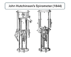
He measured the vital capacity of over 2,000 individuals. It would be a major undertaking, even in our times, to record and analyze that amount of information. He organized, prepared and published his findings in 1844. The drawings of his spirometer that accompanied his presentation are duplicated in Figure 1. He made the observation that a person’s age and height were directly related to their measured vital capacity. These facts remain unchallenged. The historic paths of Dr. Hutchinson and the vital capacity are somewhat obscure after his publication. He became quite successful in his professional life, but in 1852 he left his family and successful work in London and moved to Australia. Then he subsequently moved to Fiji where he died in 1861. His reasons for leaving England have remained unknown.
Figure 1: A reproduction of the Spirometer designed and used by Dr Hutchinson.
The history of the vital capacity and its use in lung disease is even less clear. Few techniques, procedures or therapies in medicine have clear paths of adoption. Lung diseases were among the first disorders of an organ systems to develop a focused interest from physicians. This was primarily the result of the widespread problem of pulmonary tuberculosis. The American Thoracic Society dates its origin to 1905. The use of the vital capacity measurement must have been of value to those early physicians, as by the 1940s it was being studied and a new factor was added to the standard spirometry measurements in use; the timed volumes.
In 1947 French physicians, Drs. Tiffeneau and Pinelli, using the spirometer with recording paper moving at a fixed speed, added time to the measurement of volume. One of their measurements, the forced expired volume at one second, or FEV1, has become the world standard for measuring airflow obstruction. Four years later Dr. Gaensler’s (1951) work popularized the FEV1 in the United States.
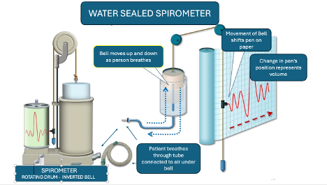
The spirometer as a testing tool has many forms. Rather than discuss the multitude of adaptations, I will focus on the water sealed model that was adapted from Dr. Hutchinson’s original design and used in the United States for most of the last half of the 20th century. Figure 2 shows a typical water sealed spirometer and its attached rotating drum with the paper recorder.
Figure 2: A standard water sealed spirometer used 1940-1980’s. The device is on the left. The container (bell) is sealed from outside air as it floats in water. Its only connection to air is through the port at the bottom right where the breathing tube (shown coiled) is attached. At the center and right of the diagram, the container (labeled bell) is shown moving up and down with the air entering and leaving it as would occur with a person breathing through the tube. It is suspended by a cord draped over a pully, at the bottom of which is a balancing weight. On the cord is a pen. As the bell moves, the pen records on paper attached to the drum next to the bell apparatus. The drum rotates at a fixed rate. The paper is removed and measurements are made with time represented along the axis of the red arrow and volume represented vertically.Diagram used by permission from: www.sibelmed.com.
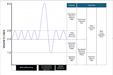
Definitions of the measurements made on the spirometer have been agreed upon and standardized for several decades. A brief review is in order. The divisions of the lung volumes as they are defined by spirometry are demonstrated in Figure 3.
Figure 3. The Lung Volumes as determined by Spirometry testing. The graph shows normal quiet breathing followed by a maximum inspiration and then by a maximum exhalation ending with normal breathing. The lung volumes are identified in the first column. The lung ‘capacities’ are identified in columns two through four. A capacity is a combination of two or three lung volumes. Image courtesy of Karri Haen Whitmer from a Mixed Course-based Research Approach to Human Physiology 2021
The rhymical up and down movement recorded initially is the air that is moved during quiet breathing. It is termed the TIDAL VOLUME.
As a deep maximal inspiration occurs, the line moves upward till no further air can be brought into the lungs. Then, as the air is exhaled, the line moves downward passing the levels of the Tidal Volume till no further air can be expelled with maximum effort. The volume of air from a maximum inhalation to a maximum exhalation is the VITAL CAPACITY, Dr. Hutchinson’s Vital Capacity.
Despite maximum effort, all the air in the lungs cannot be expelled. The amount of air that remains is termed the RESIDUAL VOLUME. Measuring this volume requires the use of additional techniques. These procedures have been well established for several decades and I will not review them unless you insist. The Residual Volume is the only volume not measured by spirometry testing. It is currently measured by one or more indirect techniques. Studies on how to measure this volume date to work by Davy in 1799.
For our understanding of the Flow Volume Loop, it is sufficient just to know that it is measured.
The addition of the Vital Capacity to the Residual Volume denotes the total amount of air the lungs contain at maximum inspiration, the TOTAL LUNG CAPACITY.
There are additional lung volumes that are very important to recognize, as breathing mechanics are different in the lung volumes above and below a certain point. At the cessation of quiet normal breathing which follows a normal Tidal Volume, there is still air left in the lung. With additional effort, some of it can be expelled till no further air will come out and only the residual volume of air remains. That additional air that can be expelled after relaxed exhalation is termed the EXPIRATORY RESERVE VOLUME.
The Expiratory Reserve Volume, when added to the Residual Volume, gives us another very important volume, the FUNCTIONAL RESIDUAL CAPACITY. The Functional Residual Capacity (FRC) is the amount of air left in the lungs at the end of relaxed breathing. To exhale additional air, effort is required. When a person exhales quietly, at the end of a tidal volume their lungs’ volume of air is the Functional Residual Capacity. It is often termed the FRC point. Above and below this point in the lung volumes the mechanics of breathing differ.
The amount of air that can be inhaled from that FRC point to maximum inhalation is termed the INSPIRATORY CAPACITY.
Spirometry measures the Tidal Volume, Vital Capacity, Inspiratory Capacity and Expiratory Reserve Volume. Knowing these volumes with the addition of a measurement of the Functional Residual Capacity, the Residual Volume can be determined.
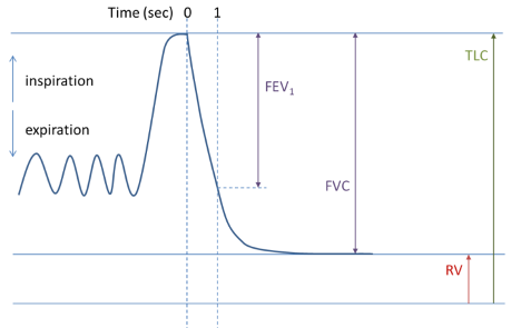
When Dr.s Tiffeneau and Pinelli added a paper drum rotating at fixed rate, timed volumes were added to the measurements. Figure 4 demonstrates the timed volume measurement of the Forced Expiratory Volume at one second (FEV1).
Figure 4. A Forced Expiratory Spirogram. On this graph, the patient inspired fully and then was instructed to blow all the air out as hard and as fast as possible. At ‘0’ time the forced exhalation has started. At the ‘1’ mark one second has passed. Note that the amount of air exhaled is the Forced Expiratory Volume at one second (FEV1.)
The FEV1 volume is the gold standard for measuring obstruction to expiratory airflow. It is expressed as a ratio to the patient’s FVC. That measurement, the FEV1 %, represents the efficiency of the patient’s ability to exhale air. Its use is worldwide.
Winston, we should next review the basic FV loop and the details of its measurement. Then a deeper look into their clinical applications and the physiology involved is needed. If you want, I could follow up with a discussion of the limitations to expiratory flow, one of my areas of interest. Yes, you can ask me to stop at any point. But assuming I don’t hear that soon, the next installment will be on its way.
Your interest in medicine outside of your specialty continues to surprise me. Yes, on a number of occasions I have mentioned the importance of the flow volume loop in testing lung functions. It has remained an interest of mine throughout my medical career. I am sure I can provide much more information than you want to know, as I was present at its birth for general use as a clinical test.
The flow volume loop is a gift to the physician. An understanding of its secrets gives the knowledgeable practitioner a world of information. It can explain or help to explain the symptoms of dyspnea and other breathing complaints. Careful review reveals more profound secrets: the unacknowledged secrets of how to understand expiratory flow, and its problems.
My love affair with the flow volume loop began in 1974 when I was made head of the pulmonary function laboratory at Walter Reed Army Medical Center. I was a newbie in the world of pulmonary physiology. Yes, I was a fully trained, knowledgeable and soon to be board certified pulmonary specialist. But the world of physiology was a whole different category of knowledge, information and details – more than any trained pulmonary physician would possess after their training in the specialty. Furthermore, I was following in the footsteps of doctors who had been more thoroughly trained in pulmonary physiology; Sidney Braman and Robert Senior, both of whom went on to extraordinary careers in academic medicine.
Drs. Hyatt and Black had just published on the value of the flow volume loop as a measurement to diagnose airflow obstructions in the trachea and larynx. The flow volume loop had not been part of our education and most patient testing facilities were not set up to make those measurements, the Walter Reed Pulmonary Testing Laboratory included. That time was before the computation of medical testing and widespread computer use in general. Flow and volume lung tests were done manually and the flow volume loop was not available to the clinician. Fortunately, or unfortunately, it is now present on almost every commercial device that measures expiratory airflow. I believe that by being so widely available, their value is being overlooked.
The technology of the 70s was evolving rapidly. Physicians were trained to understand expiratory volumes and their timing. They were also instructed in measuring basic gas changes in the blood, lung size in health and disease and the ability of gas to enter the bloodstream. These measurements were rapidly becoming common tools in the practice of clinical medicine; so too, would the new test of lung function, the flow volume loop.
Physiologists in pulmonary research had learned to measure the speed or velocity of flow with the device called a pneumotact. It provided an instant electronic signal that reflected the velocity of flowing air. Prior to that ability, volumes were measured manually and flow calculated indirectly from paper recordings. The pneumotact could be calibrated to measure the speed with which air was moving. In the 1970s this was cutting edge. Now, over 50 years later, much cheaper, more stable and reliable but similar types of devices can perform the same function and are widely available for clinical use. Almost every device currently available to measure expiratory airflow does it with some sort of electronic measurement.
The volume of air exhaled traditionally had been measured by the displacement that air would cause into a sealed container. As the air changed the position of the container, it would create a mark on paper. The inscribed marks could be physically measured and the volume of air calculated. This volume displacement was developed in the late 19th and early 20th century. In the mid 20th century, the time it took to exhale the air into the container was added as a measure of lung function. Spirometry, as it was named, had been the only pulmonary function test for measuring aspects of lung function through the 20th century and remains a mainstay of lung testing in clinical medicine today.
As a neophyte in the world of physiology, I was both frustrated and fascinated by the problems of measuring and using the flow loop. Reports by Miller and Hyatt, and then Black and Hyatt, chose to display the two parameters on an XY axis. These values needed to be measured simultaneously and plotted one verses the other. It could be projected on an oscilloscope (think of a digital screen with a line drawn on it) or on a special paper recorder. Traditionally, with spirometry testing of expiratory airflow, clinical test parameters evaluated were measurements of time on one axis and volume on the other. Those were the manually measured values. The addition of the simultaneous measurements of flow and volume, as well as the change in how they were presented, made the flow volume loop unique.
For other reasons, the lab at Walter Reed was well equipped. Previous directors had requested and received pneumotacts for use with other studies. They provided a way for me to measure the flow portion of the test. Within a few months, I was able to obtain the appropriate devices to measure volume and the special x-y recorder needed. With more than a little help from my knowledgeable staff, we created a digital signal for volume, flow, and time and then recorded them on the x-y recorder. I soon incorporated our measurement of the flow volume loop as a clinical tool. In the late 70s, subsequent upgraded testing equipment simplified these. But always in the back of my mind, those early tools brought joy to my reviews of the flow volume tests in my work.
As you can see Winston, in a relatively short period of time I was able to make the flow volume loop a clinical test available as part of the pulmonary function studies. But that was just the beginning.
Let me know if you desire more information. If you do, it may open Pandora’s flow volume box. If so, I think you would find it an interesting and valuable container.
Robert E. Hyatt MD and Black LF
Am Rev Respir Dis. 1973 Feb;107(2):191-9. doi: 10.1164/arrd.1973.107.2.191
I know it’s been a while. You haven’t heard from me in several months. Yes, I have been busy. I will try to communicate more often. First, I want you to know that all is well. Three major, well, time consuming and not ordinary events have occupied my last few months. I have completed two major projects and terminated a third.
Last summer I took extra time off from my clinical work at the Mayo. Planning ahead, I returned to it in the Fall. You will remember I was practicing part time to assist them with their backload of sleep patients. Interestingly to me, patient care is just as rewarding now as it was those years ago when I entered medicine. As they say, “You meet the nicest people.”
Computerization has ruined patient care. No, that is not exactly true. Computers have been a blessing. It is the multiple layers of regulations that are imposed through the computer programs and programmers that the doctor is burdened with completing. Large organizations are constantly modifying their systems to adjust to changing rules and regulations. I find those burdensome and onerous.
Returning to my patient care this Fall, I found it was clearly time to leave practicing clinical medicine with Mayo. My last day in September was sad but totally uneventful. So, I am officially working on my own. It is unlikely I will return to a clinical practice, but more details will follow and I will post them on the website.
“What about the other two projects?” you might wonder. They are a paper submitted and a book completed. I doubt the paper will have much success being accepted, as it is a consideration for the medical aristocracy suggesting that the AHI be modified by using total sleep time as part of the assessment for obstructive sleep apnea. I expect that I will need to offer it to another two or three journals before it is accepted for publication. Who knows, I might end up having to publish it on my own website which is certainly not the most prestigious thing to do.
And then there is the book, Reflections on Being a Physician. Depending on how I consider it, the project has lasted between three and 45 years. The book was released this year. It is a series of essays describing the trials and tribulations I associated with becoming a physician, a series about the business of medicine from a physician’s point of view and lastly, and probably most importantly, my definition or perception of the differences between a physician and a doctor. I will send you a copy. I hope you find it an interesting read. It will be available through the website at a discounted price and available through book stores and online book retailers.
One point the book stresses is the need for doctors, physicians, nurse practitioners and physician assistants, all of us who are defined as practitioners by our government, to review and recommit ourselves yearly to our oath as a provider. It is a thought you should consider.
I will get to your other questions on my next note.
You didn’t ask for this. You probably do not even want it. Yes I know, but I promised you a primer on how to apply the qualitative and quantitative analyses of insomnia complaints. Remember, insomnia as a complaint can represent many different issues. These include difficulty going to sleep and staying asleep as well as going back to sleep after awakening or combinations of those issues. Before you can apply that information, you need a simple overview of how to consider the multiple factors that can produce difficulty with sleep.
The approach I find most helpful is based on general factors known to often cause difficulty with sleep. The categories along with a brief description are:
ENVIRONMENTAL: Environmental causes are usually recognized by the patient, but not always. Factors such as pets, spouses, lighting, thermostat settings and others are some reasons people do not sleep well.
MEDICAL: At some point almost all medical conditions may interfere with sleep. Frequent conditions are usually muscular skeletal such as injuries and arthritis. Other common, though short-term, issues include allergies and upper respiratory illnesses.
MEDICATIONS: You are not surprised to see medications, are you? Their side effects on sleep is an obsession of mine. A careful review is in order. The website has a listing under the medication tab of those and of those that frequently cause excessive sleepiness and sleeplessness.
PSYCHOLOGICAL: We all experience short term stresses and emotional upheavals that lead to anxiety and depression. Long term difficulties with insomnia can result from persistent problems with anxiety and depression. Similarly, anxiety and depression can cause difficulties with sleep. The degree of anxiety and depression needed to produce these complaints is of a magnitude that should be apparent to the patient and a perceptive doctor, though psychological issues are often difficult to assess.
HABITS: Habits can produce insomnia. Familiar to all, caffeine containing products lead the list. Less well-known dietary factors are alcohol, tea and chocolate. Not for their caffeine, but for other components. The activities a person pursues prior to sleep also can play a significant role. Food, drink, exercise, TVs and computers and lighting need reviewing.
STRUCTURE: Sleep structure refers to timing of sleep. What is the patient’s sleep duration and their time spent in bed trying to sleep? How long does the patient sleep when he sleeps well? Is the patient a night owl (likes to stay up late and wake late) or a lark (likes to go to bed early and wake early)? Are the sleep hours stable or do they change on weekends. Does the patient do shift work? How much sleep does this patient actually need? All these are factors that need to be considered.
SLEEP DISORDERS: The last category of potential issues are the disorders of sleep. Recognized disorders of sleep may produce difficulty with what a patient considers insomnia. For example, obstructive sleep apnea, thought to occur in 25% of all adults, can cause difficulty with sleep maintenance but rarely causes difficulty with initiating sleep. Restless legs and periodic limb movements need to be considered when insomnia is reported. Primary insomnia, insomnia without a known cause, is a diagnosis under this group.
Applying a qualitative and quantitative assessment allows the doctor to focus on specific areas of concern. Often, a provider will consider a patient to have primary insomnia without giving the other factors consideration. They take the patient’s complaints of insomnia – the patient’s perceptions of their sleep difficulties as insomnia – to be insomnia without a cause and proceed with medications to help induce sleep. Getting needed information from the patient is often difficult and time can limit the providers’ opportunities to explore other possible issues. For example, frequently a patient will tell me they have stopped using caffeinated products, but upon questioning, tea or energy drinks are still being used. Another major issue is medications. Patients may be on multiple medications which can produce sleeplessness or sleepiness. It can be impossible at times to separate their specific effects.Clearly identifying the details of the complaint will direct the provider to specific items to be considered for the patients diagnosis and treatment.
It is possible to identify contributing or causative factors in many patients with insomnia complaints. The qualitative and quantitative assessments often lead to specific addressable issues that can be modified or treated without the use of sleep-inducing medications. My working patterns can be summarized best in a tabular form that I have reproduced below.
When a patient’s complaints fall into one of these patterns, a more detailed history and investigation to assess that issue will follow.
And yes, Winston, I will try not to bother you with any more advice on insomnia.
RGH 29430
COMMON PATTERNS OF INSOMNIA COMPLAINTS IN SPECIFIC SLEEP PROBLEMS