
Winston
I know you are aware of the basics of lung terminology and testing, but it is important that the terms be fresh in your mind before I answer your questions about the Flow Volume Loop and why I thought it was so important to review them.
Hutchinson was a surgeon who recognized that the ability to breathe deeply was important to a person’s health. He created the device used for the next century and a half to measure breathing and named it the spirometer. He then proceeded to measure the maximum amount of air that a person could inspire and he termed it the Vital Capacity. His definition of Vital Capacity is used to this day. The spirometer, while changed in shape, appearance and measuring methods, is still the name used for devices that measure the vital capacity as well as other lung parameters.
You will not be surprised to learn that many of the great names in history, including medical history, had an interest in the lungs and their function: Individuals such as Aristotle, Galen, Harvey, Toricelli and Hooke. The list goes on. In the narrow field of lung function testing, the names are not going to be familiar. They will include John Hutchinson (1844), Tiffeneau with Pinelli (1947) and Gaensler (1951). Despite listing their names and giving these gentlemen recognition, they represent just a small percentage of the hundreds and probably thousands of doctors who added to the early understanding of lung functions and testing.
So what is a spirometer? Dr. Hutchinson instructed individuals to breathe through a tube connected to a closed container that could expand and contract as air entered and left it. To achieve this in the 1800s, he used an upsidedown can as the container which was suspended in and sealed from other air by water. The movement of the container was measured and the amount of air the person was able to exhale was calculated. The concept was adapted and used widely throughout the mid 20th century.
He measured the vital capacity of over 2,000 individuals. It would be a major undertaking, even in our times, to record and analyze that amount of information. He organized, prepared and published his findings in 1844. The drawings of his spirometer that accompanied his presentation are duplicated in Figure 1. He made the observation that a person’s age and height were directly related to their measured vital capacity. These facts remain unchallenged. The historic paths of Dr. Hutchinson and the vital capacity are somewhat obscure after his publication. He became quite successful in his professional life, but in 1852 he left his family and successful work in London and moved to Australia. Then he subsequently moved to Fiji where he died in 1861. His reasons for leaving England have remained unknown.
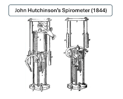
Figure 1: A reproduction of the Spirometer designed and used by Dr Hutchinson.
The history of the vital capacity and its use in lung disease is even less clear. Few techniques, procedures or therapies in medicine have clear paths of adoption. Lung diseases were among the first disorders of an organ systems to develop a focused interest from physicians. This was primarily the result of the widespread problem of pulmonary tuberculosis. The American Thoracic Society dates its origin to 1905. The use of the vital capacity measurement must have been of value to those early physicians, as by the 1940s it was being studied and a new factor was added to the standard spirometry measurements in use; the timed volumes.
In 1947 French physicians, Drs. Tiffeneau and Pinelli, using the spirometer with recording paper moving at a fixed speed, added time to the measurement of volume. One of their measurements, the forced expired volume at one second, or FEV1, has become the world standard for measuring airflow obstruction. Four years later Dr. Gaensler’s (1951) work popularized the FEV1 in the United States.
The spirometer as a testing tool has many forms. Rather than discuss the multitude of adaptations, I will focus on the water sealed model that was adapted from Dr. Hutchinson’s original design and used in the United States for most of the last half of the 20th century. Figure 2 shows a typical water sealed spirometer and its attached rotating drum with the paper recorder.
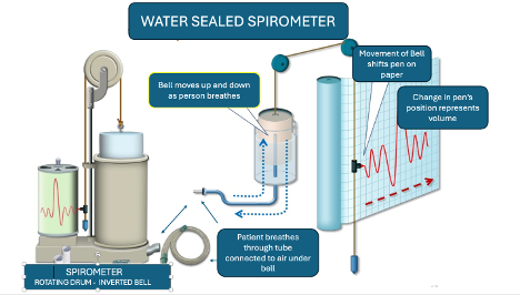
Figure 2: A standard water sealed spirometer used 1940-1980’s. The device is on the left. The container (bell) is sealed from outside air as it floats in water. Its only connection to air is through the port at the bottom right where the breathing tube (shown coiled) is attached. At the center and right of the diagram, the container (labeled bell) is shown moving up and down with the air entering and leaving it as would occur with a person breathing through the tube. It is suspended by a cord draped over a pully, at the bottom of which is a balancing weight. On the cord is a pen. As the bell moves, the pen records on paper attached to the drum next to the bell apparatus. The drum rotates at a fixed rate. The paper is removed and measurements are made with time represented along the axis of the red arrow and volume represented vertically.Diagram used by permission from: www.sibelmed.com.
Definitions of the measurements made on the spirometer have been agreed upon and standardized for several decades. A brief review is in order. The divisions of the lung volumes as they are defined by spirometry are demonstrated in Figure 3.
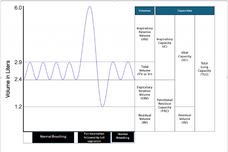
Figure 3. The Lung Volumes as determined by Spirometry testing. The graph shows normal quiet breathing followed by a maximum inspiration and then by a maximum exhalation ending with normal breathing. The lung volumes are identified in the first column. The lung ‘capacities’ are identified in columns two through four. A capacity is a combination of two or three lung volumes. Image courtesy of Karri Haen Whitmer from a Mixed Course-based Research Approach to Human Physiology 2021
The rhymical up and down movement recorded initially is the air that is moved during quiet breathing. It is termed the TIDAL VOLUME.
As a deep maximal inspiration occurs, the line moves upward till no further air can be brought into the lungs. Then, as the air is exhaled, the line moves downward passing the levels of the Tidal Volume till no further air can be expelled with maximum effort. The volume of air from a maximum inhalation to a maximum exhalation is the VITAL CAPACITY, Dr. Hutchinson’s Vital Capacity.
Despite maximum effort, all the air in the lungs cannot be expelled. The amount of air that remains is termed the RESIDUAL VOLUME. Measuring this volume requires the use of additional techniques. These procedures have been well established for several decades and I will not review them unless you insist. The Residual Volume is the only volume not measured by spirometry testing. It is currently measured by one or more indirect techniques. Studies on how to measure this volume date to work by Davy in 1799.
For our understanding of the Flow Volume Loop, it is sufficient just to know that it is measured.
The addition of the Vital Capacity to the Residual Volume denotes the total amount of air the lungs contain at maximum inspiration, the TOTAL LUNG CAPACITY.
There are additional lung volumes that are very important to recognize, as breathing mechanics are different in the lung volumes above and below a certain point. At the cessation of quiet normal breathing which follows a normal Tidal Volume, there is still air left in the lung. With additional effort, some of it can be expelled till no further air will come out and only the residual volume of air remains. That additional air that can be expelled after relaxed exhalation is termed the EXPIRATORY RESERVE VOLUME.
The Expiratory Reserve Volume, when added to the Residual Volume, gives us another very important volume, the FUNCTIONAL RESIDUAL CAPACITY. The Functional Residual Capacity (FRC) is the amount of air left in the lungs at the end of relaxed breathing. To exhale additional air, effort is required. When a person exhales quietly, at the end of a tidal volume their lungs’ volume of air is the Functional Residual Capacity. It is often termed the FRC point. Above and below this point in the lung volumes the mechanics of breathing differ.
The amount of air that can be inhaled from that FRC point to maximum inhalation is termed the INSPIRATORY CAPACITY.
Spirometry measures the Tidal Volume, Vital Capacity, Inspiratory Capacity and Expiratory Reserve Volume. Knowing these volumes with the addition of a measurement of the Functional Residual Capacity, the Residual Volume can be determined.
When Dr.s Tiffeneau and Pinelli added a paper drum rotating at fixed rate, timed volumes were added to the measurements. Figure 4 demonstrates the timed volume measurement of the Forced Expiratory Volume at one second (FEV1).
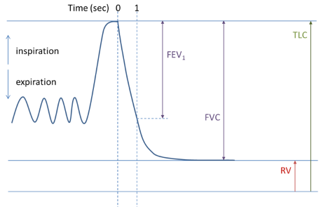
Figure 4. A Forced Expiratory Spirogram. On this graph, the patient inspired fully and then was instructed to blow all the air out as hard and as fast as possible. At ‘0’ time the forced exhalation has started. At the ‘1’ mark one second has passed. Note that the amount of air exhaled is the Forced Expiratory Volume at one second (FEV1.)
The FEV1 volume is the gold standard for measuring obstruction to expiratory airflow. It is expressed as a ratio to the patient’s FVC. That measurement, the FEV1 %, represents the efficiency of the patient’s ability to exhale air. Its use is worldwide.
Winston, we should next review the basic FV loop and the details of its measurement. Then a deeper look into their clinical applications and the physiology involved is needed. If you want, I could follow up with a discussion of the limitations to expiratory flow, one of my areas of interest. Yes, you can ask me to stop at any point. But assuming I don’t hear that soon, the next installment will be on its way.
0 Comments